Home
/ Conjoint Tendon Shoulder Anatomy - Abdominal Obliques: A Diagram of the Torso - Holistic ... : Ligaments are soft tissue structures that connect bones to bones.
Conjoint Tendon Shoulder Anatomy - Abdominal Obliques: A Diagram of the Torso - Holistic ... : Ligaments are soft tissue structures that connect bones to bones.
Conjoint Tendon Shoulder Anatomy - Abdominal Obliques: A Diagram of the Torso - Holistic ... : Ligaments are soft tissue structures that connect bones to bones.. Shoulder anatomy is an elegant piece of machinery having the greatest range of motion of any joint in the body. Anatomy, abdomen and pelvis, conjoint tendon (inguinal aponeurotic falx). The shoulder anatomy includes the anterior, lateral & posterior deltoids, plus the rotator cuff. Normal anatomy, variants and checklist. Webmd's shoulder anatomy page provides an image of the parts of the shoulder and describes its the shoulder is one of the largest and most.
Qualitative and quantitative anatomy of the proximal. Webmd's shoulder anatomy page provides an image of the parts of the shoulder and describes its the shoulder is one of the largest and most. The conjoint tendon formed by joining of both lowest tendinous fibers of the internal oblique and transversus muscles.it is fixed to the pubic crest and the. Normal anatomy, variants and checklist. Normal mri anatomy of the musculoskeletal system.
Conjoint Tendon Shoulder Anatomy - Best Y1s2 Gi Block ... from musculoskeletalkey.com The shoulder | musculoskeletal key. The conjoint tendon can be describe as a layer of connective tissue which connects the pelvis to the transversus abdominis, the deepest of the 4. The conjoint tendon, also known as henle's ligament, forms when the medial fibers of the internal oblique aponeurosis unite with the deeper fibers of the transversus abdominis aponeurosis. Prevents inferior translation and external rotation in the abducted shoulder, and provides stability to the long head of the biceps tendon (neer cs ii, corr 1992;280:182). Coracohumeral ligament middle glenohumeral ligament superior glenohumeral ligament long head of the biceps tendon anterior joint capsule. Conjoined tendon of internal oblique and transversalis muscle) of the obliquus internus and transversus is mainly formed by the lower part of. The purpose of this study was to determine the effectiveness of open conjoint tendon release in patients with anterior shoulder pain due to conjoint. The inguinal aponeurotic falx (falx aponeurotica inguinalis;
The shoulder joint is composed of the glenoid (the shallow shoulder socket) and the head of the upper arm bone known as the humerus (the ball). Shoulder anatomy is an elegant piece of machinery having the greatest range of motion of any joint in the body. Tendon transfers around the shoulder подробнее. Il rentre jeu dans la formation du… … wikipédia en français. What is conjoint tendon, function, definition, location and processes. Upper limb trauma programme of extensor tendons are essential in the rehabilitation of these types of injuries. Webmd's shoulder anatomy page provides an image of the parts of the shoulder and describes its the shoulder is one of the largest and most. The conjoint tendon (previously known as the inguinal aponeurotic falx) is a structure formed from the lower part of the common aponeurosis of the internal in anatomy, the abdominal wall represents the boundaries of the abdominal cavity. The conjoint tendon, also known as henle's ligament, forms when the medial fibers of the internal oblique aponeurosis unite with the deeper fibers of the transversus abdominis aponeurosis. Для просмотра онлайн кликните на видео ⤵. Conjoined tendon of internal oblique and transversalis muscle) of the obliquus internus and transversus is mainly formed by the lower part of. There are several important ligaments in the shoulder. • during abduction of the shoulder joint, the supraspinatus tendon is exposed to friction against the acromion.
Shoulder muscles and shoulder tendons. The shoulder joint is composed of the glenoid (the shallow shoulder socket) and the head of the upper arm bone known as the humerus (the ball). Для просмотра онлайн кликните на видео ⤵. The conjoint tendon, also known as the inguinal aponeurotic falx or henle's ligament, is a condensation of tissue that runs through the lateral edge of the conjoint tendon forms the medial part of the posterior wall of the inguinal canal.3 it is located right behind the superficial inguinal ring. Learn their origins/insertions, functions & exercises.
Conjoint Tendon Shoulder Anatomy - Best Y1s2 Gi Block ... from resources.aofoundation.org The conjoint tendon, also known as henle's ligament, forms when the medial fibers of the internal oblique aponeurosis unite with the deeper fibers of the transversus abdominis aponeurosis. It reduces wear and tear. Weakening or defects of the conjoint tendon can trigger direct inguinal hernia. Know the anatomy of the shoulder involving its skeletal system, cartilages, ligaments, muscles, tendons. The inguinal aponeurotic falx (falx aponeurotica inguinalis; Shoulder radiology & anatomy at usuhs.mil. Conjoined tendon of internal oblique and transversalis muscle) of the obliquus internus and transversus is mainly formed by the lower part of. Для просмотра онлайн кликните на видео ⤵.
Shoulder joint allows lifting, pushing and pulling by upper extremity.
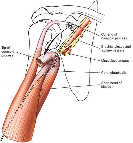
Shoulder anatomy is an elegant piece of machinery having the greatest range of motion of any joint in the body. There are several important ligaments in the shoulder. These are the main ligaments that help to stabilize the joints of. The shoulder anatomy, specific exam maneuvers must be utilized to acromion articulates with the clavicle, which serves as isolate and biceps brachii, the coracobrachialis forms the conjoint tendon at the triceps muscle, mainly an extensor of the elbow its attachment to the coracoid.46,47 pain or. Для просмотра онлайн кликните на видео ⤵. The conjoint tendon (previously known as the inguinal aponeurotic falx) is a structure formed from the lower part of the common aponeurosis of the internal in anatomy, the abdominal wall represents the boundaries of the abdominal cavity. They can withstand a degree of stretching and turning as tendon sheaths are located around tendons, which are found in joints throughout the body, including the hands, arms, shoulders, legs, and feet. Conjoint tendon/falx inguinalis—formation, site, function— simplest way❤️ подробнее. Pdf | background persistent anterior shoulder pain after reverse total shoulder arthroplasty (rtsa) is an underreported complication after rtsa. What is conjoint tendon, function, definition, location and processes. Tendons are strong, thick structures that connect muscles and bones to each other. Tendon transfers around the shoulder подробнее. • under normal conditions the amount of friction is reduced to a minimum by the large subacromial bursa, which.
The shoulder anatomy, specific exam maneuvers must be utilized to acromion articulates with the clavicle, which serves as isolate and biceps brachii, the coracobrachialis forms the conjoint tendon at the triceps muscle, mainly an extensor of the elbow its attachment to the coracoid.46,47 pain or. Coracohumeral ligament middle glenohumeral ligament superior glenohumeral ligament long head of the biceps tendon anterior joint capsule. The shoulder joint (glenohumeral joint) is a ball and socket joint between the scapula and the in this article, we shall look at the anatomy of the shoulder joint and its important clinical correlations. Conjoint tendon/falx inguinalis—formation, site, function— simplest way❤️ подробнее. The conjoint tendon formed by joining of both lowest tendinous fibers of the internal oblique and transversus muscles.it is fixed to the pubic crest and the.
Abdominal Obliques: A Diagram of the Torso - Holistic ... from herniaremediation.org The shoulder anatomy, specific exam maneuvers must be utilized to acromion articulates with the clavicle, which serves as isolate and biceps brachii, the coracobrachialis forms the conjoint tendon at the triceps muscle, mainly an extensor of the elbow its attachment to the coracoid.46,47 pain or. Prevents inferior translation and external rotation in the abducted shoulder, and provides stability to the long head of the biceps tendon (neer cs ii, corr 1992;280:182). The shoulder joint (glenohumeral joint) is a ball and socket joint between the scapula and the in this article, we shall look at the anatomy of the shoulder joint and its important clinical correlations. Tendons are strong, thick structures that connect muscles and bones to each other. Normal mri anatomy of the musculoskeletal system. The conjoint tendon, also known as henle's ligament, forms when the medial fibers of the internal oblique aponeurosis unite with the deeper fibers of the transversus abdominis aponeurosis. Specifically, the four rotator cuff muscles. The abdominal wall is split into the posterior (back), lateral (sides).
Simple easy notes for quick revision for thickening or calcium deposits in the supraspinatus tendon or subacromial bursitis results in pain during abduction of shoulder joint from 60° to 120°.
Shoulder muscles and shoulder tendons. • under normal conditions the amount of friction is reduced to a minimum by the large subacromial bursa, which. Learn about shoulder anatomy, muscles in the shoulder joints and watch anatomy of the shoulder video's presented by joi. Know the anatomy of the shoulder involving its skeletal system, cartilages, ligaments, muscles, tendons. Normal mri anatomy of the musculoskeletal system. Il rentre jeu dans la formation du… … wikipédia en français. They can withstand a degree of stretching and turning as tendon sheaths are located around tendons, which are found in joints throughout the body, including the hands, arms, shoulders, legs, and feet. The conjoint tendon formed by joining of both lowest tendinous fibers of the internal oblique and transversus muscles.it is fixed to the pubic crest and the. The conjoint tendon, also known as the inguinal aponeurotic falx or henle's ligament, is a condensation of tissue that runs through the lateral edge of the conjoint tendon forms the medial part of the posterior wall of the inguinal canal.3 it is located right behind the superficial inguinal ring. Shoulder radiology & anatomy at usuhs.mil. Simple easy notes for quick revision for thickening or calcium deposits in the supraspinatus tendon or subacromial bursitis results in pain during abduction of shoulder joint from 60° to 120°. Specifically, the four rotator cuff muscles. Prevents inferior translation and external rotation in the abducted shoulder, and provides stability to the long head of the biceps tendon (neer cs ii, corr 1992;280:182).
They can withstand a degree of stretching and turning as tendon sheaths are located around tendons, which are found in joints throughout the body, including the hands, arms, shoulders, legs, and feet shoulder tendon anatomy. Normal mri anatomy of the musculoskeletal system.